The post Beyond Average: New Benchmarks for Excellence in Pediatric Emergency Care appeared first on ALiEM.
]]>
DOI: 10.1001/jamanetworkopen.2026.13391 | PubMed: PMID 42154467
Emergency departments routinely track quality measures: door-to-provider time, length of stay, return visits, medication administration, and dozens of others. However, knowing your performance is only half the equation. The harder question is what benchmark should you be aiming for?
Comparing with the national average may tell you where you stand, but “average” is not necessarily synonymous with good care. At the other extreme, setting every target at 100% adherence or zero failures may be unrealistic, and can direct substantial resources toward marginal gains.
A recent study by Alpern and colleagues in JAMA Network Open offers a different approach [1]. Using more than 5.3 million pediatric emergency department visits from the Pediatric Emergency Care Applied Research Network (PECARN) Registry, the investigators established Achievable Benchmarks of Care (ABCs) for common measures of pediatric emergency care. Rather than defining excellence theoretically, they asked a pragmatic question: What level of performance are the best-performing clinicians already achieving in real-world emergency departments?
The Study
The investigators performed a cross-sectional analysis of 5,302,587 visits from 2017 through 2024 across 12 emergency departments participating in the PECARN Registry: nine pediatric EDs and three affiliated community sites. The analysis included 925 clinicians. PECARN has also published a brief summary of the study [2].
The authors evaluated measures spanning several important domains of pediatric emergency care:
- Initial assessment and vital-sign documentation
- Timeliness and ED throughput
- Return visits and other quality and safety measures
- Asthma care
- Pain assessment and treatment
- Antibiotic stewardship
Many of these are familiar quality metrics already endorsed or prioritized by organizations involved in pediatric emergency care. The important innovation was not the measures themselves. It was how the target for each measure was established.
What Is an “Achievable Benchmark”?
Instead of using the mean or median as the standard, the investigators identified the highest-performing clinicians who collectively cared for approximately 10% of eligible visits. Performance among this high-achieving group was then used to calculate the benchmark.
For binary measures, the investigators used an adjusted performance fraction designed to prevent clinicians with small numbers of eligible encounters from disproportionately influencing the benchmark. Continuous measures were ranked using median performance. The result is a target grounded in achievable excellence. The distinction matters. Whereas the mean tells an ED what everyone else is doing, an ABC provides evidence of what high-performing clinicians are already capable of doing under real clinical conditions.
The table below shows selected measures from a condensed list provided by the study’s lead author, chosen as the most important or interesting targets for change.
| Performance measure | Achievable benchmark |
|---|---|
| ED flow (throughput) | |
| Time to provider, all patients | 20 min |
| ESI 2 | 13 min |
| ESI 3 | 19 min |
| ESI 4 | 22 min |
| ED length of stay, all patients | 114 min |
| Discharged | 110 min |
| Admitted or transferred | 268 min |
| Clinical care | |
| Systemic corticosteroids for asthma | 97.8% |
| Time to first beta-agonist for asthma | 29 min |
| ≥2-point pain reduction with long-bone fracture | 80.5% |
| After the visit | |
| Return visit within 48 hours | 1.6% |
Where Is the Opportunity to Improve?
Performance was not uniform across sites. For example, the proportion of children with long-bone fractures achieving at least a 2-point reduction in pain ranged from 61.4% to 92.6% across sites. Systemic corticosteroid administration for asthma ranged from 93.1% to 99.2%. Vital-sign documentation also showed substantial variation. In contrast, return visits resulting in admission and antibiotic stewardship showed relatively little variation.
That pattern may offer important direction for quality improvement. Measures that are relatively close to the clinician’s locus of control, such as asthma treatment and fracture pain management, showed meaningful differences between average site performance and what the site’s best-performing clinicians could achieve. That changes the quality-improvement conversation. If clinicians in the same ED, caring for similar patients within the same system, are consistently achieving better performance, the question becomes less “Can we do this?” and more “What are they doing differently, and how can we make that the default?”
Throughput Is Different
Door-to-provider time and length of stay deserve more cautious consideration. The study found substantial variation in throughput benchmarks between sites and between pediatric EDs and affiliated community settings. Interestingly, throughput performance worsened over the study period.
Unlike giving corticosteroids for an asthma exacerbation, ED length of stay is only partly controlled by an individual clinician. Crowding, staffing, boarding, diagnostic resources, consultation availability, inpatient capacity, and patient volume all can influence throughput. The authors appropriately note that future benchmarks may need to account for volume and capacity.
For example, a 110-minute benchmark for a discharged pediatric patient should not automatically become an inflexible performance mandate for every ED. It is better interpreted as a reference point: this level of performance has been demonstrated, and a substantial gap from it should prompt examination of the systems contributing to that gap.
Why Benchmarking Against “Average” Isn’t Enough
Quality dashboards frequently compare a clinician or department with an institutional or national mean. That approach is intuitive but has an obvious weakness: the average describes current performance; it does not necessarily define desirable performance.
Imagine an ED with a median time to provider of 38 minutes. Learning that the network average is 36 minutes tells the department that it is slightly slower than its peers. Learning that high-performing clinicians and systems can achieve approximately 20 minutes asks a much more useful question: what would have to change to close that gap?
The same concept applies to clinical care. An asthma corticosteroid benchmark of 97.8% is not an aspirational number invented by a committee. It reflects performance that clinicians in actual emergency departments have demonstrated.
ABCs therefore occupy a useful space between two imperfect targets:
“Be average” ← Achievable excellence → “Be perfect.”
A Benchmark Is a Starting Point, not a Report Card
There are important limitations. The PECARN Registry sites are predominantly pediatric centers, along with affiliated community sites. They do not represent the general EDs that care for most children nationally. Differences in pediatric volume, staffing, resources, geography, and pediatric readiness may affect whether a particular benchmark is immediately attainable in another setting. The study also relies on electronic health record data. Documentation does not always represent care, and EHR-derived measures cannot capture every element of clinical judgment or patient outcome.
These benchmarks therefore should not be interpreted as universal thresholds separating “good” from “bad” EDs, or good from bad clinicians. They are something more useful: empirical targets showing what high-performing clinicians and systems have demonstrated is possible.
The Bottom Line
Emergency medicine has no shortage of quality measures. What has often been missing is a meaningful answer to the question, “What should our target be?” This article provides one approach. By deriving benchmarks from high performers across more than 5.3 million pediatric ED encounters, the study team moves the target away from both average performance and theoretical perfection and toward achievable excellence.
For clinicians and ED leaders, the most useful question may not be whether your department meets every benchmark tomorrow. Instead, look at the measures where your performance differs most from what high-performing clinicians have already demonstrated. Determine which gaps are driven by individual practice and which are driven by systems. Then ask: What is keeping excellent care from becoming our usual care?
References
- Alpern ER, Ramgopal S, Olsen CS, et al; PECARN Registry Study Group. Achievable benchmarks of care in pediatric emergency departments. JAMA Netw Open. 2026;9(5):e2613391. PMID: 42154467. doi:10.1001/jamanetworkopen.2026.13391
- Woods JM. What excellent care looks like: new benchmarks. Pediatric Emergency Care Applied Research Network (PECARN). Published June 23, 2026. https://pecarn.org/pecarn_news/what-excellent-care-looks-like-new-benchmarks/
Author information

The post Beyond Average: New Benchmarks for Excellence in Pediatric Emergency Care appeared first on ALiEM.
]]>The post ALiEM AIR Series | Ortho Spine Module (2026) appeared first on ALiEM.
]]>
Welcome to the AIR Ortho Spine Module! After carefully reviewing all relevant posts in the past 11 months from the top 50 sites of the Digital Impact Factor, the ALiEM AIR Team is proud to present the highest quality online content related to orthopedic spine emergencies in the Emergency Department. 6 blog posts met our standard of online excellence and were approved for residency training by the AIR Series Board. More specifically, we identified 2 AIR and 4 Honorable Mentions. We recommend programs give 3 hours of III credit for this module.
AIR Stamp of Approval and Honorable Mentions
In an effort to truly emphasize the highest quality posts, we have 2 subsets of recommended resources. The AIR stamp of approval is awarded only to posts scoring above a strict scoring cut-off of ≥30 points (out of 35 total), based on our scoring instrument. The other subset is for “Honorable Mention” posts. These posts have been flagged by and agreed upon by AIR Board members as worthwhile, accurate, unbiased, and appropriately referenced despite an average score.
Want asynchronous Individualized Interactive Instruction (III) credit?
Take the AIR quiz at ALiEMU. Free, 1-time login required.
Highlighted Quality Posts: Ortho Spine 2026
| Site | Article | Author | Date | Label |
|---|---|---|---|---|
| EMCrit | Spinal Cord | Dr. Josh Farkas | March 4, 2026 | AIR |
| EMCrit | Spinal Epidural Abscess (SEA) | Dr. Josh Farkas | January 5, 2026 | AIR |
| emDocs | EM@3AM: Cervical Myelopathy | Drs. Derick Sanchez and Michael Cirone | January 10, 2026 | HM |
| St Emlyn’s Blog | Early Blood Pressure Targets in Acute Spinal Cord Injury | Dr. Simon Carley | January 17, 2026 | HM |
| The SGEM | SGEM#483: Electricity – TENS Units for Treating Back Pain | Dr. Wm. Ken Milne | August 23, 2025 | HM |
| RCEMLearning | Spinal Injuries in Children | Dr. Liz Herrieven | July 29, 2025 | HM |
(AIR = Approved Instructional Resource; HM = Honorable Mention)
If you have any questions or comments on the AIR series, or this AIR module, please contact us!
Author information

The post ALiEM AIR Series | Ortho Spine Module (2026) appeared first on ALiEM.
]]>The post Pediatric Pneumonia Without Routine Chest X-Rays: How Much Weight Should We Give the Lung Exam? appeared first on ALiEM.
]]>
DOI: 10.1001/jamanetworkopen.2026.30074 | PubMed: PMID 42623068
Have you stood at the bedside while the resident, nurse, and student each described a different lung examination? Someone heard focal crackles. Someone else heard coarse breath sounds. The next clinician heard clear lungs. The PECARN PedCAPS (Pediatric Community-Acquired Pneumonia Severity) study suggests that this familiar disagreement is common.
In a new PECARN PedCAPS analysis published in JAMA Network Open, two clinicians independently examined the same 252 children with community-acquired pneumonia (CAP). No individual physical examination finding met the study’s prespecified reliability standard [1]. A single lung sound should not decide the diagnosis of pneumonia.
This creates a clinical dilemma.
The Pediatric Infectious Diseases Society (PIDS)/Infectious Diseases Society of America (IDSA) guidelines advise against routine chest radiography to confirm suspected CAP in children well enough for outpatient treatment [2]. However, the clinician must still decide whether CAP is likely enough to treat. That decision depends heavily on the history and physical examination, even though no individual symptom or sign is sufficiently sensitive or specific and clinicians may interpret the same lung sounds differently [3].
How do we then diagnose outpatient CAP without reflexively ordering an x-ray?
Study Design
This planned reliability analysis was nested within the ongoing prospective PECARN PedCAPS study cohort. Investigators enrolled children ages 3 months through 17 years with clinician-diagnosed CAP at seven academic pediatric EDs. Two attending physicians, advanced practice clinicians, or PEM fellows independently examined each child within 60 minutes. The clinicians completed identical standardized forms but received no special training in how to define the examination findings.
The study population limits how far we can take the results. Nearly all participants, 249 of 252 (98.8%), had pneumonia supported by chest radiography, and 128 (50.8%) were hospitalized. This selected group had established CAP and differs from the undifferentiated population of children presenting with cough, fever, or respiratory distress.
Investigators measured raw agreement and Fleiss κ for the following:
- Altered mental status
- Capillary refill
- Grunting
- Retractions or accessory muscle use
- Auscultatory lung findings:
- Wheezing
- Decreased breath sounds
- Crackles or rales
- Rhonchi or coarse breath sounds
Acceptable reliability required the lower bound of the κ 95% confidence interval to be at least 0.40.
Results
No finding passed the prespecified threshold. Wheezing and retractions performed best, but only reached moderate agreement:
- Wheezing: κ = 0.50 (95% CI 0.39-0.62), with 82% raw agreement
- Retractions or accessory muscle use: κ = 0.49 (95% CI 0.37-0.60), with 78% raw agreement
- Crackles or rales: κ = 0.21 (95% CI 0.11-0.31), with 55% raw agreement
- Rhonchi or coarse breath sounds: κ = 0.18 (95% CI 0.08-0.28), with 56% raw agreement
- Decreased breath sounds: κ = 0.11 (95% CI 0.02-0.20), with 46% raw agreement
When wheezing was simplified to present or absent without asking clinicians to distinguish focal from diffuse findings, κ improved to 0.58. This suggests that localization added another layer of disagreement.
Reliability did not differ significantly between the 124 children discharged home and the 128 hospitalized. The analyses by age, race, and clinician knowledge of chest radiograph results were underpowered and should not be described as showing equivalence across those groups.
Altered mental status, delayed capillary refill, and grunting were uncommon. Their high overall raw agreement largely reflected agreement that the findings were absent. When at least one examiner documented one of these findings, agreement that it was present was low. Clinicians should still treat altered mental status and grunting as concerning when present.
How to Diagnose Without a Routine Chest X-Ray?
Reliability and diagnostic accuracy are different questions. This study shows that clinicians frequently disagree about examination findings in children already diagnosed with CAP. It did not measure the sensitivity, specificity, or likelihood ratios of those findings among all children with possible pneumonia. Because almost every participant had radiographic pneumonia, it also cannot determine how auscultation performs in the less selected population we evaluate on shift.
The PIDS/IDSA guideline discourages routine imaging while preserving selective imaging when uncertainty matters. It does, however, recommend a chest radiograph when a child has suspected or documented hypoxemia, significant respiratory distress, or failure of initial antibiotic therapy [2]. In a well-appearing child without those features, a clinician should holistically combine the history with measured respiratory rate, oxygen saturation, work of breathing, overall appearance, serial examinations, and a reliable follow-up plan. One reported crackle, focal decrease in breath sounds, or coarse sound should not become the diagnostic push for radiographs.
“Our results emphasize the need to look at multiple features of the clinical presentation rather than exam findings in isolation when making the diagnosis of pneumonia.”
Todd A. Florin, MD, MSCE, senior author of this 2026 JAMA publication
Bottom Line
Keep listening to the lungs, but do not let one auscultatory finding decide whether a child has pneumonia. When examinations conflict and the uncertainty would change management, return to the full clinical picture and obtain additional information selectively.
References
- Hooli S, Reeder R, Cutler L, et al. Reliability of Physical Examination Findings in Youths Diagnosed With Pneumonia. JAMA Netw Open. 2026;9(8):e2630074. PMID: 42623068. doi:10.1001/jamanetworkopen.2026.30074
- Bradley JS, Byington CL, Shah SS, et al. The Management of Community-Acquired Pneumonia in Infants and Children Older Than 3 Months of Age: Clinical Practice Guidelines by the Pediatric Infectious Diseases Society and the Infectious Diseases Society of America. Clin Infect Dis. 2011;53(7):e25-e76. PMID: 21880587. doi:10.1093/cid/cir531
- Lipsett SC, Neuman MI. Rethinking Auscultation for Pediatric Pneumonia: Do My Ears Deceive Me? JAMA Netw Open. 2026;9(8):e2630083. PMID: 42623079. doi:10.1001/jamanetworkopen.2026.30083
Author information

The post Pediatric Pneumonia Without Routine Chest X-Rays: How Much Weight Should We Give the Lung Exam? appeared first on ALiEM.
]]>The post Trick of the Trade: A Fast Exit Strategy for the TACO Method in Heat Stroke Resuscitation appeared first on ALiEM.
]]>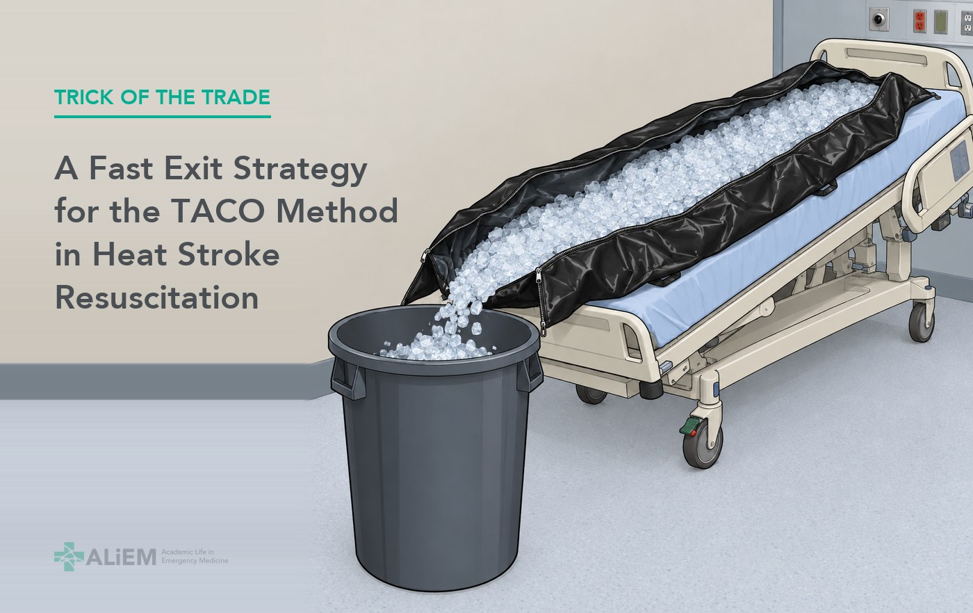
An unidentified male presents to the emergency department via ambulance after being found unresponsive in the heat. His initial rectal temperature is 108°F (42.2°C), and his GCS is 4 for nondescript groaning. The team quickly secures the airway and places a temperature-sensing urinary catheter while ice arrives from the cafeteria. They start rapid cooling, the cornerstone of heat stroke management, with the Tarp-Assisted Cooling with Oscillation (TACO) method and reach 102°F (38.9°C) quickly [1,2]. After a brief sigh of relief, another problem arises: how do you rapidly stop the cooling process to avoid overshooting into hypothermia?
The team now faces several logistical challenges in this common but time-sensitive scenario, including the management of multiple points of unwieldy but critical tubing while moving the patient, minimizing spillage of contaminated water, and, most importantly, preventing hypothermia. Most cooling protocols focus on how to start cooling by immersion, and not so much on how to end it. Our quick and simple trick adds one vital tool: gravity.
Trick of the Trade
Tilt the bed foot-down, cut open the foot of the body bag, and let gravity drain the ice-water slurry into a trash bin.
Materials
- Disposable body bag, used as the TACO immersion vessel
- Large trash bin
- Trauma shears
- Towels
Steps
- Perform TACO in a disposable body bag. While cooling is underway, place a large trash bin at the foot of the bed.
- Once core temperature reaches 102°F (38.9°C), raise the bed to maximum height.
- Place the bed in reverse Trendelenburg, so the foot of the bed tilts down.
- Use trauma shears to cut across the foot of the body bag. Be careful of the patient’s toes!
- Have the team push the ice-water slurry into the bin, with gravity to assist.
- When most of the ice water is in the bin, towel-dry the patient.
In our case, the patient was dry in less than 2 minutes and never became hypothermic. The team could then remove the body bag and reposition the patient safely, without the time pressure.
Tips
- Time-saving maneuver: if the patient needs an airway before cooling, place the temperature-sensing urinary catheter while intubation is underway.
- Continuous core temperature monitoring allows early recognition of when to stop active cooling.
- Reverse Trendelenburg lets gravity assist with rapid evacuation. This buys time later to reposition the patient carefully and avoid accidental removal of critical lines and tubes.
Read more from the Tricks of the Trade series.
References
- Luhring KE, Butts CL, Smith CR, et al. Cooling effectiveness of a modified cold-water immersion method after exercise-induced hyperthermia. J Athl Train. 2016;51(11):946-951. doi: 10.4085/1062-6050-51.12.07
- Comp G, Pugsley P, Sklar D, et al. Heat stroke management updates: a description of the development of a novel in-emergency department cold-water immersion protocol and guide for implementation. Ann Emerg Med. 2025;85(1):43-52. doi: 10.1016/j.annemergmed.2024.07.013
Author information

The post Trick of the Trade: A Fast Exit Strategy for the TACO Method in Heat Stroke Resuscitation appeared first on ALiEM.
]]>The post Sickle Cell Acute Pain Episodes: Lessons Learned from the Prematurely Halted PECARN STArT Trial appeared first on ALiEM.
]]>
DOI: 10.1001/jama.2026.13310 | PubMed: PMID 42616542
A child with sickle cell disease arrives in your emergency department (ED) in an acute pain episode. In one of the largest US pediatric trials of a treatment for acute sickle cell pain, how long that crisis lasted varied far more between hospitals than it did between the study drug and placebo.
The PECARN STArT trial randomized 274 children and young adults with sickle cell acute pain episodes to IV arginine or saline placebo at 10 US children’s hospitals. Arginine, which targets the nitric oxide depletion that drives vaso-occlusion, did not shorten time to crisis resolution, and the trial was halted early for futility [1]. The more useful numbers are in site-level data. Median time to crisis resolution varied by as much as 61 hours between participating hospitals, and mean total parenteral opioid by as much as 3.0 mg/kg of based on morphine-equivalent calculations [1,3]. These are observational site differences. They may reflect analgesia and opioid-discontinuation practice, patient mix, and other institutional factors that the analyses can not distinguish.
Study Design
STArT was a double-blind, placebo-controlled phase 3 randomized clinical trial conducted (2021-2024). Patients aged 3 to 21 years with sickle cell disease who presented to the ED with acute pain requiring parenteral opioids and hospital admission were randomized within 12 hours of their first IV opioid dose. Arginine was given as a 200 mg/kg loading dose followed by 100 mg/kg every 8 hours until discharge. Of 274 patients randomized, 271 received study drug, 129 arginine and 142 placebo. The primary outcome was time to crisis resolution, the interval from first study drug delivery to the last dose of parenteral opioid, and it is the outcome the US Food and Drug Administration prefers for regulatory approval in this population [1].
Results
Median time to crisis resolution was 60.8 hours with arginine versus 65.8 hours with placebo, an absolute difference of 7.2 hours favoring arginine with a confidence interval spanning benefit in either direction (95% CI, -21.6 to 35.9 hours). Total parenteral opioid use, pain scores, and hospital length of stay were similar between groups, and the arginine effect on time to crisis resolution did not differ significantly by chronic pain status. Serious adverse events did not differ and there were no deaths [1].
Three descriptive findings arose from the collected data:
- Site variation. Median time to crisis resolution differed by up to 61 hours across the 10 hospitals, and mean total parenteral opioid by up to 3.0 mg/kg [1,3].
- Delay to study drug. Study drug arrived a median of 8 hours after the first ED dose of IV opioid, and only 50.4% of participants had sought care within 24 hours of pain onset [1]. In 23 of 271 treated participants the last IV opioid had already been given before study drug arrived, producing a primary outcome of zero hours.
- Chronic pain prevalence. 41% of participants met criteria for chronic sickle cell pain, including 34% of children younger than 12 years [1]. A single triage pain score means something different in a child who already has pain 15 or more days a month.
Build an Expedited Pain Plan in the Emergency Department
It is unclear why the wide variation in time to pain crisis resolution. The study was not designed to untangle the reasons, but in theory the causes are likely multifactorial and include: patient mix, underlying chronic pain, time spent in pain before arrival, institutional practices, and individual practitioner analgesia practices. That last one is something that you can act on today.
An expedited pain plan for a sickle cell acute pain episode
Consider administering intranasal fentanyl at triage, if IV opioids are not readily available.
The American Society of Hematology 2020 recommendation is to assess the patient and administer analgesia within 1 hour of ED arrival, with reassessment every 30 to 60 minutes. It is a strong recommendation on low-certainty evidence [8].
Two cross-sectional PECARN studies describe what meeting those targets looks like. In a PECARN Registry analysis of 9233 ED visits for uncomplicated sickle cell pain at 12 children’s hospitals, a first opioid dose within 60 minutes of arrival was associated with lower odds of hospitalization (OR 0.84; 95% CI, 0.75-0.95). That association held even when the second dose fell outside 30 minutes (OR 0.85), and with a timely first dose the odds fell stepwise as the second-dose interval shortened: 0.78 within 60 minutes, 0.70 within 45, and 0.62 within 30 (95% CI, 0.52-0.75) [9]. Separately, among 400 children at 20 academic pediatric EDs in the US and Canada, only 19% received intranasal fentanyl, and those who did had nearly ninefold higher adjusted odds of discharge from the ED (adjusted OR 8.99; 95% CI, 2.81-30.56; P < .001) [10].
Both studies are cross-sectional and neither establishes causality. Timeliness of the first dose is the more robust of the two signals, since it survived a late second dose.
Not All Pain is the Same
The STArT study enrolled acute pain episodes, defined by pain severe enough to need parenteral opioids. However, arginine specifically targets vaso-occlusion pain. The accompanying JAMA editorial argued that these are not the same thing: the term acute pain episode has become operationally synonymous with vaso-occlusive episode, yet acute pain in sickle cell disease also arises from neuropathic mechanisms, central sensitization, and musculoskeletal injury [2]. A trial that heterogeneously enrolls all of them dilutes the study population. Notably, 41% of the study participants had underlying chronic pain [1].
Can We Narrow the Patient Inclusion Criteria?
The editorial accompanying STArT argues for enrolling a more narrow subset of patients, focusing on objective markers rather than pain severity [2]:
- Hypoxemia, worrisome for acute chest syndrome
- Very high LDH with thrombocytopenia, reported in a small retrospective adult series as suggesting a severe vaso-occlusive subtype that prompted consideration of early intervention [6]
- Acute kidney injury or acute liver injury, used the way organ dysfunction defines severity in sepsis trials
Bottom Line
- Arginine is not ready. Nothing in this STArT trial displaces supportive care, NSAIDs, and opioids for a sickle cell acute pain episode.
- The 61-hour spread between hospitals was more than three times the trial’s originally hypothesized 17-hour treatment difference. How your department assesses and treats pain is part of what that spread reflects.
- The American Society of Hematology recommends giving an analgesic dose within the first hour of ED arrival. Consider intranasal fentanyl at triage, while awaiting IV access.
References
- Morris CR, Hatabah D, Korman R, et al; Pediatric Emergency Care Applied Research Network (PECARN). Arginine therapy for sickle cell disease acute pain episodes: the STArT randomized clinical trial. JAMA. Published online August 19, 2026. PMID: 42616542. doi:10.1001/jama.2026.13310
- Anum SJ, Kanter J. Arginine treatment and sickle cell disease pain: a great STArT, but a hard end point. JAMA. Published online August 19, 2026. PMID: 42616535. doi:10.1001/jama.2026.14849
- Rees CA, Hatabah D, Korman R, et al; PECARN. Hospital variations in time-to-crisis-resolution among children and adolescents with sickle cell disease. Am J Hematol. 2026;101(1):206-212. PMID: 41190764. doi:10.1002/ajh.70129
- Morris CR, Ahmad F, Airewele G, et al. Sickle cell disease treatment with arginine therapy (STArT): results of a phase-3 randomized controlled trial. Blood. 2025;146(suppl 1):616. doi:10.1182/blood-2025-616
- Brousseau DC, Scott JP, Badaki-Makun O, et al. A multicenter randomized controlled trial of intravenous magnesium for sickle cell pain crisis in children. Blood. 2015;126(14):1651-1657. PMID: 26232172. doi:10.1182/blood-2015-05-647107
- Gardner K, Thein SL. Super-elevated LDH and thrombocytopenia are markers of a severe subtype of vaso-occlusive crisis in sickle cell disease. Am J Hematol. 2015;90(10):E206-E207. PMID: 26205137. doi:10.1002/ajh.24126
- Ataga KI. The challenge of clinical end points in sickle cell disease. Blood. 2023;142(24):2047-2054. PMID: 37890140. doi:10.1182/blood.2023021220
- Brandow AM, Carroll CP, Creary S, et al. American Society of Hematology 2020 guidelines for sickle cell disease: management of acute and chronic pain. Blood Adv. 2020;4(12):2656-2701. PMID: 32559294. doi:10.1182/bloodadvances.2020001851
- Gwarzo I, Coleman KD, McKinley K, et al. Opioid timeliness in the emergency department and hospitalizations for acute sickle cell pain. JAMA Pediatr. 2025;179(11):1194-1202. PMID: 40892426. doi:10.1001/jamapediatrics.2025.2967
- Rees CA, Brousseau DC, Ahmad FA, et al; SCD Arginine Study Group and PECARN. Intranasal fentanyl and discharge from the emergency department among children with sickle cell disease and vaso-occlusive pain: a multicenter pediatric emergency medicine perspective. Am J Hematol. 2023;98(4):620-627. PMID: 36606705. doi:10.1002/ajh.26837
- Gladwin MT, Kato GJ, Weiner D, et al; DeNOVO Investigators. Nitric oxide for inhalation in the acute treatment of sickle cell pain crisis: a randomized controlled trial. JAMA. 2011;305(9):893-902. PMID: 21364138. doi:10.1001/jama.2011.235
- Casella JF, Barton BA, Kanter J, et al. Effect of poloxamer 188 vs placebo on painful vaso-occlusive episodes in children and adults with sickle cell disease: a randomized clinical trial. JAMA. 2021;325(15):1513-1523. PMID: 33877274. doi:10.1001/jama.2021.3414
- Dampier CD, Telen MJ, Wun T, et al; RESET Investigators. A randomized clinical trial of the efficacy and safety of rivipansel for sickle cell vaso-occlusive crisis. Blood. 2023;141(2):168-179. PMID: 35981565. doi:10.1182/blood.2022015797
Author information
The post Sickle Cell Acute Pain Episodes: Lessons Learned from the Prematurely Halted PECARN STArT Trial appeared first on ALiEM.
]]>The post Trick of the Trade: Improvised Collar and Cuff Sling Using Soft Limb Holders appeared first on ALiEM.
]]>
A 69-year-old woman presents to the emergency department (ED) after 2 falls at home. She tripped on a rug overnight, felt dizzy as she tried to get up, and fell again. She struck her head but has no pain from it. Her chief complaint is right shoulder pain, and she reports frequent shoulder dislocations in the past. An x-ray shows an acute comminuted, non-displaced fracture of the humeral neck with involvement of the greater tuberosity.
Initial management is immobilization in a sling for 1-3 weeks, and the evidence supports nonoperative treatment for most proximal humerus fractures [1,2]. A collar and cuff sling is an appropriate management plan to allow the elbow to hang free, so gravity applies gentle traction that helps maintain fracture alignment. However, what if your ED does not stock such a sling?
Trick of the Trade
Build a collar and cuff sling from 2 Posey soft limb holders, the padded wrist restraints most EDs already stock. In our case, this improvised collar and cuff sling provided adequate support to the wrist and a cushion for the patient’s neck.
Materials
2 Posey soft limb holders:
- Use one complete holder.
- Use only the padding from the second holder.
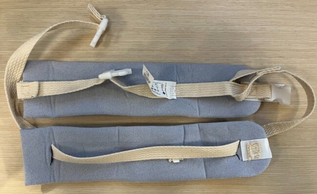
Top padding from full Posey holder for the wrist; bottom padding from second Posey holder for neck collar
Steps
- Wrap one cuff around the wrist of the injured arm.
- Remove the padding from the second cuff, and thread that padding onto the long strap of the first cuff.
- Pass the strap behind the patient’s neck. Slide the padding along the strap until it cushions the back of the neck. This is the collar.
- Adjust the strap length so the elbow rests at 90 degrees with the wrist slightly above the elbow, and secure the buckle.
- Improvised collar and cuff sling made from a Posey soft limb holder, worn with the elbow at 90 degrees and the wrist supported
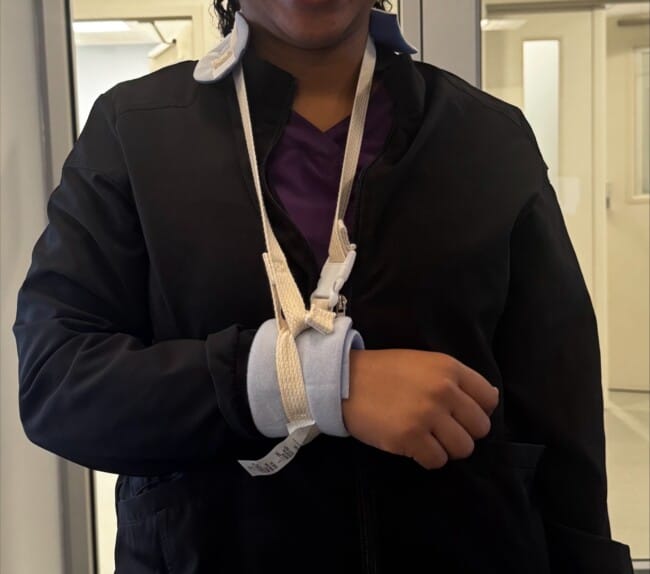
Improvised collar and cuff setup
Tips
- Application of the padded neck collar is important, because the Posey strap is often thin, flat, and coarse. It can dig into the neck once it carries the weight of an arm over the next 1-3 weeks.
- Support the wrist only and allow the elbow to hang to gravity. This axial traction helps to maintain fracture alignment.
Interested in More?
For a collar and cuff improvised with the neck strap from a shoulder immobilizer, plus other common ED splinting techniques, see SplintER Series: Common ED Splint Techniques 104.
References
- Handoll HH, Elliott J, Thillemann TM, Aluko P, Brorson S. Interventions for treating proximal humeral fractures in adults. Cochrane Database Syst Rev. 2022;6(6):CD000434. doi: 10.1002/14651858.CD000434.pub5
- Rangan A, Handoll H, Brealey S, et al. Surgical vs nonsurgical treatment of adults with displaced fractures of the proximal humerus: the PROFHER randomized clinical trial. JAMA. 2015;313(10):1037-1047. doi: 10.1001/jama.2015.1629
Author information

The post Trick of the Trade: Improvised Collar and Cuff Sling Using Soft Limb Holders appeared first on ALiEM.
]]>The post ALiEM AIR Series | ACS Cardiology Module (2026) appeared first on ALiEM.
]]>
Welcome to the AIR ACS Cardiology Module! After carefully reviewing all relevant posts in the past 11 months from the top 50 sites of the Digital Impact Factor [1], the ALiEM AIR Team is proud to present the highest quality online content related to ACS cardiology emergencies in the Emergency Department. 5 blog posts met our standard of online excellence and were approved for residency training by the AIR Series Board. More specifically, we identified 1 AIR and 4 Honorable Mentions. We recommend programs give 3 hours of III credit for this module.
AIR Stamp of Approval and Honorable Mentions
In an effort to truly emphasize the highest quality posts, we have 2 subsets of recommended resources. The AIR stamp of approval is awarded only to posts scoring above a strict scoring cut-off of ≥30 points (out of 35 total), based on our scoring instrument. The other subset is for “Honorable Mention” posts. These posts have been flagged by and agreed upon by AIR Board members as worthwhile, accurate, unbiased, and appropriately referenced despite an average score.
Want asynchronous Individualized Interactive Instruction (III) credit?
Take the AIR quiz at ALiEMU. Free, 1-time login required.
Highlighted Quality Posts: ACS Cardiology 2026
| Site | Article | Author | Date | Label |
|---|---|---|---|---|
| EM Crit | 2025 AHA and ESICM guidelines on post-arrest care | Dr. Josh Farkas | October 26, 2025 | AIR |
| EM Crit | Type-1 MI (OMI and NOMI) and related complications | Dr. Josh Farkas | May 1, 2025 | HM |
| The Bottom Line | MINT – Restrictive or Liberal transfusion strategy in MI | Dr. Daniel Chung | May 23, 2025 | HM |
| EM Crit | Impella Management | Dr. Josh Farkas | October 14, 2024 | HM |
| EM Crit | ST elevation | Dr. Josh Farkas | November 5, 2024 | HM |
(AIR = Approved Instructional Resource; HM = Honorable Mention)
If you have any questions or comments on the AIR series, or this AIR module, please contact us!
Author information
The post ALiEM AIR Series | ACS Cardiology Module (2026) appeared first on ALiEM.
]]>The post PERC-Peds Rule Could Change How Children Are Evaluated for Pulmonary Embolism: A PECARN Study appeared first on ALiEM.
]]>
DOI: 10.1016/S2213-2600(26)00086-X
Pulmonary embolism (PE) in children is uncommon, but delayed or missed diagnosis can have serious consequences. Despite this, there is relatively little evidence to guide clinician evaluation of suspected pediatric PE. There are no validated pediatric PE rule-out strategies and adult diagnostic pathways have not been prospectively tested in children.
PECARN sought to change that with their recently published study – BEEPER: Bedside Exclusion of Pulmonary Embolism without Radiation in Children [1]. The investigators developed a rule that safely excluded PE in low-risk children.
Study Objective
The goal of BEEPER was to prospectively evaluate whether the Pulmonary Embolism Rule-out Criteria adapted for children (PERC-Peds) could safely exclude pulmonary embolism in children without the need for laboratory testing or imaging.
What is PERC-Peds?
PERC-Peds was adapted from the adult Pulmonary Embolism Rule-out Criteria (PERC), a well-established adult clinical decision rule used to exclude PE in low-risk patients without additional testing [2]. Using retrospective pediatric data, investigators modified the adult PERC rule to create the pediatric version. A child was considered PERC-Peds negative only if ALL of the following were true:
- Clinician gestalt pretest probability <15%
- No prior PE or proximal DVT
- No surgery requiring intubation within 30 days
- No current estrogen use
- No hemoptysis
- Heart rate always:
- <100 beats/min if older than 12 years
- <120 beats/min if 12 years or younger
- Oxygen saturation consistently >94%
- No suspected DVT
Study Design
A Large, Prospective, Multicenter PECARN Study
BEEPER was a multicenter, prospective observational diagnostic accuracy study conducted across 21 emergency departments within PECARN (the Pediatric Emergency Care Applied Research Network).
Inclusion Criteria:
- 4–17 years old, AND
- had suspected PE or proximal DVT prompting clinicians to:
- order diagnostic testing, OR
- strongly consider PE in the differential diagnosis
Eligible diagnostic testing included:
- D-dimer
- CT pulmonary angiography (CTPA)
- V/Q scan
- MRI angiography
- Other pulmonary vascular imaging
All testing decisions remained entirely at clinician discretion.
Outcome Definition
The outcome was venous thromboembolism (VTE), including:
- image-confirmed pulmonary embolism, OR
- proximal DVT (above knee or elbow)
Results
Enrollment
- 4,039 children enrolled, 3,988 had analyzable data
Prevalence of Disease
Overall:
- 254 children (6.3%) had PE and/or proximal DVT
Breakdown:
- Isolated proximal DVT: 76
- Both PE and proximal DVT: 56
- Isolated PE: 122
Performance of PERC-Peds
The PERC-Peds rule demonstrated extremely high diagnostic sensitivity.
Diagnostic Accuracy
- Sensitivity: 99.6% (95% CI 97.8–99.9%)
- Specificity: 19.6% (95% CI 18.4–21.0%)
- False negative rate: 0.1% (95% CI 0–0.75%)
- Negative predictive value: 99.9%
There was only ONE false negative. These findings suggest that PERC-Peds can safely exclude PE in a subset of low-risk children without additional laboratory testing or imaging.
D-Dimer Findings
BEEPER also provided the first large prospective evaluation of D-dimer performance in children undergoing PE evaluation. Clinicians ordered D-dimer testing in approximately 75–79% of enrolled children. Using a standardized threshold of 500 ng/mL:
- Sensitivity: 88.8%
- Specificity: 61.9%
- False negative rate: 1.2–1.4%
These data support the use of D-dimer as part of a sequential diagnostic strategy in pediatric PE evaluation for those children who fail PERC-Peds.
PERC-Peds + D-Dimer Sequential Strategy
One of the most clinically important findings was the potential value of combining:
- PERC-Peds
- followed by D-dimer if PERC-Peds failed
This sequential strategy would have:
- safely excluded PE in 54.7% of enrolled children,
- excluded PE in 69% of children who had D-dimer ordered,
- maintained an acceptable false negative rate of 1.0% (95% CI 0.6–1.5%),
- estimated reduction in CT pulmonary angiography use by approximately 18.5%.
Putting PERC-Peds in Context
The PERC-Peds rule performed well compared to the regularly utilized adult PERC rule. Adult PERC studies [2] typically show sensitivities around 95% with false negative rates below 2%, while in BEEPER, the sensitivity approached 100% with an exceptionally low false negative rate. Just as importantly, this is the first prospective study to support a bedside rule-out strategy for PE in children: offering a potential pathway to safely reduce unnecessary CT scans and radiation exposure in low-risk patients. BEEPER also lays the groundwork for a more standardized pediatric PE evaluation approach, where clinicians could use PERC-Peds first, followed by D-dimer testing when needed, reserving imaging for children at higher risk.
What’s Next?
While BEEPER represents a major step forward in pediatric PE diagnosis, the study also highlights important limitations and unanswered questions. PERC-Peds demonstrated high sensitivity, but relatively low specificity: meaning many children will still fail the rule, and indiscriminate use could potentially increase testing. Importantly, BEEPER was an observational diagnostic study, meaning it did not evaluate outcomes from the implementation of a clinical decision rule.
BEEPER provides the foundation for a new era of pediatric PE evaluation research. Future studies will likely focus on implementation science: understanding whether use of PERC-Peds changes clinician behavior, safely reduces imaging, and can be integrated into real-world emergency department workflows. Investigators also envision future diagnostic algorithms that combine PERC-Peds with D-dimer testing to reserve imaging for children at highest risk, potentially reducing unnecessary radiation exposure in a meaningful number of patients. Additional work can evaluate performance across different clinical settings, refine the gestalt component of the rule, and explore integration into electronic health record decision support tools. Taken together, BEEPER fills one of the most important evidence gaps in pediatric emergency medicine and represents the first major step toward safer, more standardized, and evidence-based pediatric PE evaluation.
References
- Ellison AM, Kuppermann N, Shihabuddin BS, et al. PERC-Peds rule for bedside exclusion of pulmonary embolism without radiation in children in the USA (BEEPER): a multicentre, prospective, observational, diagnostic accuracy study. Lancet Respir Med. Published online July 2026. doi:10.1016/S2213-2600(26)00086-X.
- Kline JA, Courtney DM, Kabrhel C, et al. Prospective multicenter evaluation of the pulmonary embolism rule-out criteria. J Thromb Haemost. 2008;6(5):772-780. doi:10.1111/j.1538-7836.2008.02944.x. PMID: 18318689.
Author information

The post PERC-Peds Rule Could Change How Children Are Evaluated for Pulmonary Embolism: A PECARN Study appeared first on ALiEM.
]]>The post What We Still Do Not Know About Pediatric Mental Health Emergencies: PECARN Research Agenda appeared first on ALiEM.
]]>
DOI: 10.1016/j.annemergmed.2026.05.015 | PubMed: PMID 42429726
Every emergency physician knows these moments. A 10-year-old with autism is escalating in a hallway bed, and nobody can say which de-escalation approach or which medication is safest for him. A 15-year-old is boarding for a third night after a suicide attempt, receiving no active treatment while she waits for a psychiatric bed. A charge nurse asks whether universal suicide screening is worth the workflow disruption. These are routine clinical decisions, and for most of them the pediatric evidence simply does not exist.
A new consensus statement from the PECARN Mental Health Working Group, A Research Agenda for Acute Pediatric Mental and Behavioral Health Emergencies, published in Annals of Emergency Medicine, maps where those evidence gaps are and which ones matter most [1]. The panel reached consensus on 51 research priorities, 31 of them top tier. Read as a whole, the agenda is an unusually honest inventory of how much of current pediatric behavioral health emergency care runs on extrapolation and local habit.
Background
One in 6 US children has a mental or behavioral health condition, and nearly half receive no treatment from a mental health professional [2]. The ED has become the de facto safety net: visits for self-harm and harm to others have risen substantially over the past decade, boarding times have stretched, and in 2021 the American Academy of Pediatrics, the American Academy of Child and Adolescent Psychiatry, and the Children’s Hospital Association jointly declared a national emergency in child and adolescent mental health [3]. What has not kept pace is the evidence for what emergency clinicians should actually do during these visits.
How the Agenda Was Built
The working group used a modified Delphi process with 23 expert partners: 4 parents and 1 young adult with lived experience of pediatric mental health emergency care, general and pediatric emergency physicians, emergency nurses, child and adolescent psychiatrists with emergency expertise, ED social workers, out-of-hospital and EMS-fellowship-trained clinicians, and a research funder. Across 3 survey rounds, 76 literature-informed candidate priorities were modified, expanded, and rated on need and urgency, research impact, and family centeredness. The prespecified retention criteria required agreement from both the family representative group and the clinician and funder group, so family voices could not be outvoted. The result: 51 consensus priorities sorted into 3 tiers.
Where the Gaps Are
Suicide prevention dominates
Suicide prevention questions account for 42% of the top tier. The panel wants to know which suicide risk screening tool best predicts real outcomes such as return visits, attempts, and deaths, including in neurodivergent children and non-English speakers; what universal screening does to those outcomes; and whether safety planning, with or without structured follow-up calls, actually increases mental health follow-up and reduces future attempts in youth. The contrast with adult evidence is striking. In adults, the multicenter ED-SAFE trial showed that universal screening plus a brief intervention reduced post-discharge suicidal behavior [4]. The pediatric equivalent has never been done. The panel also prioritized a practical lethal-means question: which safety devices, from medication lock boxes to firearm safes, do caregivers actually prefer and use after the ED visit?
Agitation: everything is still an open question
Which de-escalation methods best reduce medication use, restraint use, and staff injuries, and how does that differ by age, developmental stage, culture, language, and trauma history? Which medications are safest and most effective, for which children? What works for children with neurodevelopmental disorders such as autism spectrum disorder, who face a higher risk of pharmacologic and physical restraint? None of these have comparative evidence today, even though documented racial disparities in restraint application make the stakes plain [1].
Boarding and ED care
Top-tier questions include whether brief therapy delivered in the ED during boarding improves symptoms, shortens length of stay, and lowers admission rates, and whether home-based care is a safe and acceptable alternative to hospitalization. The panel also flagged care in rural and low-resource EDs, where most of these children are actually seen.
Before and after the ED
Out-of-hospital priorities center on training first responders in de-escalation and trauma-informed care and on testing novel response models, including mental health co-response teams and alternative destinations. A single-county pilot of direct EMS transport to a psychiatric emergency facility found that roughly 2 in 5 encounters met criteria for direct transport, with only 0.5% requiring secondary ED transfer within 24 hours [5]. On the back end, the agenda targets the high-risk post-discharge window: what actually gets youth to mental health care after they leave, and whether stepped-care models using telehealth reduce return visits and future attempts.
Clinical Implications
Nothing in a research agenda changes tomorrow’s orders, and this post will not pretend otherwise. The value for a practicing clinician is different. First, the agenda names how thin the floor is under common practices: medication choice for acute agitation in children, for example, is largely extrapolated from adult psychiatry. Knowing where evidence is absent should make us slower to treat local protocol as settled science. Second, this document signals where PECARN and federal funders will direct pediatric emergency mental health research over the next 5 to 10 years, which is useful for anyone building a QI program, a research career, or a departmental protocol they would prefer not to rewrite twice. Third, the top-tier questions double as an audit checklist: if your ED cannot say how often it restrains children, whether safety planning happens before discharge, or what its mental health boarding times are, the agenda is a reasonable place to start measuring.
One limitation deserves mention because the authors themselves flag it: no expert partners identified as Black or Hispanic, a notable gap given the documented racial disparities in restraint use and in behavioral health triage that PECARN’s own work has described.
Bottom Line
PECARN’s consensus research agenda distills the pediatric mental and behavioral health emergency evidence gap into 51 prioritized questions, with suicide prevention accounting for 42% of the top tier. It will not change your practice today. It tells you something more uncomfortable and more useful: for most of what we do during pediatric behavioral health visits, from de-escalation to safety planning to boarding care, the evidence base has not been written yet, and this is the field’s official to-do list for writing it.
References
- Hoffmann JA, Foster AA, Krass P, et al. A research agenda for acute pediatric mental and behavioral health emergencies. Ann Emerg Med. Published online July 10, 2026. PMID: 42429726. doi:10.1016/j.annemergmed.2026.05.015
- Bitsko RH, Claussen AH, Lichstein J, et al. Mental health surveillance among children – United States, 2013-2019. MMWR Suppl. 2022;71(2):1-42. PMID: 35202359. doi:10.15585/mmwr.su7102a1
- American Academy of Pediatrics, American Academy of Child and Adolescent Psychiatry, Children’s Hospital Association. AAP-AACAP-CHA declaration of a national emergency in child and adolescent mental health. 2021.
- Miller IW, Camargo CA Jr, Arias SA, et al. Suicide prevention in an emergency department population: the ED-SAFE study. JAMA Psychiatry. 2017;74(6):563-570. PMID: 28456130. doi:10.1001/jamapsychiatry.2017.0678
- Glomb NW, Trivedi T, Grupp-Phelan J, et al. Safety of a prehospital emergency medical services protocol for an alternative destination for pediatric behavioral emergencies in Alameda County. J Am Coll Emerg Physicians Open. 2023;4(2):e12930. PMID: 37051504. doi:10.1002/emp2.12930
Author information
The post What We Still Do Not Know About Pediatric Mental Health Emergencies: PECARN Research Agenda appeared first on ALiEM.
]]>The post ACMT Toxicology Visual Pearl – The Fang and the Furious appeared first on ALiEM.
]]>
Envenomation from this oceanic snake would most likely lead to which of the following symptoms?
- Acute renal failure
- Blindness
- Descending paralysis
- Localized skin necrosis
[Image courtesy of Petr Hamernik – Zoo Praha, Wikimedia Commons]

The American College of Medical Toxicology hosts this Toxicology Visual Pearls series. The post was peer-reviewed on behalf of ACMT by Drs Spencer Greene, Howard A Greller, and Louise Kao.
References
- Falla MV, Sousa EP, Morais-Zani K, et al. Functional and Proteomic Characterization of Acanthophis Antarcticus Venom: Evidence of Fibrinogenolytic and Serine Peptidase Inhibitory Activities. Toxins (Basel). 2025;17(8):405. PMID: 40864081.
- Fry BG, Wickramaratna JC, Jones A, Alewood PF, Hodgson WC. Species and Regional Variations in the Effectiveness of Antivenom Against the in Vitro Neurotoxicity of Death Adder (Acanthophis) Venoms. Toxicol Appl Pharmacol. 2001;175(2):140-148. PMID: 11543646.
- Tasoulis T, Wang CR, Ellis S, et al. The Venom Proteome of the Ecologically Divergent Australian Elapid, Southern Death Adder Acanthophis Antarcticus. Toxins (Basel). 2025;17(7):352. PMID: 40711163.
- Johnston CI, O’Leary MA, Brown SG, et al. Death Adder Envenoming Causes Neurotoxicity Not Reversed by Antivenom–Australian Snakebite Project (ASP-16). PLoS Negl Trop Dis. 2012;6(9):e1841. PMID: 23029595.
- Blacklow B, Konstantakopoulos N, Hodgson WC, Nicholson GM. Presence of Presynaptic Neurotoxin Complexes in the Venoms of Australo-Papuan Death Adders (Acanthophis Spp.). Toxicon. 2010;55(6):1171-1180. PMID: 20064542.
- Currie BJ. Snakebite in Tropical Australia: A Prospective Study in the “Top End” of the Northern Territory. Med J Aust. 2004;181(11-12):693-697. PMID: 15588215.
- Seifert SA, Armitage JO, Sanchez EE. Snake Envenomation. N Engl J Med. 2022;386(1):68-78. PMID: 34986287.
- Lieu K, Livshits Z, LeSaint KT. Venomous Snakes and Snakebites. JAMA. 2025;334(16):1494. PMID: 40875216.
- Blacklow B, Escoubas P, Nicholson GM. Characterisation of the Heterotrimeric Presynaptic Phospholipase A(2) Neurotoxin Complex From the Venom of the Common Death Adder (Acanthophis Antarcticus). Biochem Pharmacol. 2010;80(2):277-287. PMID: 20361942.
- Isbister GK, Brown SG, Page CB, et al. Snakebite in Australia: A Practical Approach to Diagnosis and Treatment. Med J Aust. 2013;199(11):763-768. PMID: 24329653.
- Lalloo DG, Trevett AJ, Black J, et al. Neurotoxicity, Anticoagulant Activity and Evidence of Rhabdomyolysis in Patients Bitten by Death Adders (Acanthophis Sp.) in Southern Papua New Guinea. QJM. 1996;89(1):25-35. PMID: 8730340.
- Tibballs J. Australian Snake Antivenom Dosing: What Is Scientific and Safe? Anaesth Intensive Care. 2020;48(2):129-133. PMID: 31505950.
- Warrell DA, Williams DJ. Clinical Aspects of Snakebite Envenoming and Its Treatment in Low-Resource Settings. Lancet. 2023;401(10385):1382-1398. doi:10.1016/S0140-6736(23)00002-8. PMID: 36931290.
- Flachsenberger W, Mirtschin P. Anticholinesterases as Antidotes to Envenomation of Rats by the Death Adder (Acanthophis Antarcticus). Toxicon. 1994;32(1):35-39. doi:10.1016/0041-0101(94)90019-1. PMID: 9237335.
Author information

The post ACMT Toxicology Visual Pearl – The Fang and the Furious appeared first on ALiEM.
]]>