When: Monday, November 11, 6:30 – 8:30 p.m.
Where: Hilton San Diego Bayfront, 1 Park Blvd, San Diego, CA 92101
Room: Sapphire 400
Moderator:
- Noah Gray, Nature
Panel:
- Susanna-Assunta Sansone, Assoc Dir & PI at University of Oxford e-Research Centre; Honorary Academic Editor, Scientific Data
- Mark Hahnel, Figshare
- Kalyani Narasimhan, Nature Neuroscience
- Sean Hill, International Neuroinformatics Coordinating Facility (INCF)
- Maryann Martone, University of California San Diego, UCSD (Neuroscience Information Framework)
Plus after the panel discussion join us for a networking event where our panellists will be joined by:
- Brian Wandell (Stanford): fMRI data
- Laura Colgin (U. Texas): Navigational behavior/neural spike data
- Andrew Hires (USC): Microscope imaging/sensorimotor behavior data
Please tweet any questions for the panel using the hastag #SfN13data
Places are limited and only 150 delegates will be admitted.
BACK GROUND:
Tackling the terabyte: how should research adapt to the era of big data?
It’s getting easier and easier to collect gigabytes of data, but many scientists lack the skills to use it or are trained in how to best organize it.
Computer models also produce very complex theoretical predictions, but how do you compare the outcome of a complex model with the original data? How much duplicated data exists? What rights management is associated with data reuse? And should data that’s made available for reuse be peer-reviewed?
Managing such large volumes of data represents a fundamental change in how experimental science is done, is the research community prepared for this change? How should we start to tackle this problem? Should universities have data specialists? And who would pay for them? Aren’t we all busy enough already?
Other topics to consider:
- Will journals act as depository for ‘Big Data’?
- Neuroinformatics – integration/analysis of large-volume, high-dimensional & fine-grain experimental data (Neuroscience Database Gateway)
- Where do we go with neurosci data – (driving policy, adding value to papers, author credit) ?
- Mandate the sharing of information?
- Should all research be peer reviewed – Blogs vs social media?
- Using social networking to aid research (collaboration)
- What should ‘journals’ (Independent v Society) be doing to facilitate / How can journals help?

- Original image courtesy of Stuart Miles / FreeDigitalPhotos.net

We’re back! Apologies for the long radio silence – day job, what can I say.
Last week Nature published a leader reflecting upon our performance as editors and journalists in the gender balance of our referees, commissioned authors, and journalistic profiles. The verdict? Plenty of room for improvement – in 2011, only 14% of Nature’s 5,514 manuscript referees were women. Those numbers are for all areas, both physical and life sciences. I don’t have the exact number for just neuroscientists but a quick partial analysis suggests it is in the same ballpark. How good/bad is 14%? According to a 2007 survey of North American neuroscience programs, 36% of neuroscience assistant professors, 28% of associate, and 21% of full professors are women. I don’t know what those percentages would be if you included neuroscientists from the rest of the the world (I’m guessing they would be lower), but I am fairly confident in saying we haven’t been grossly overrepresenting women in our referee picks.
So how do we choose our referees? In a nutshell: for each paper, we pick 3 (sometimes 2, sometimes more) people with expertise in the topic of the manuscript who we know to be rigorous, but fair, and timely reviewers. We try to ensure that all technical aspects of the study are covered, so sometimes we will bring people in who may not be experts in the subfield, but can assess individual experimental techniques used in the study. Most of the time we pick people who have reviewed for us before, not just because we think they are good reviewers, but so that we can appropriately weight their reviews based on our previous experiences of their editorial toughness, general stances on standards for their field, etc. We sometimes incorporate reviewer suggestions made by the authors if the suggested people seem appropriate to us, and if we already have experience with them. We don’t send papers to people excluded by the authors (within reason), and we try not to send papers to people’s bosom buddies, mortal enemies, recent collaborators or mentors (to the best of our knowledge). Newer referees are balanced with familiar ones on the roster so that we have a sense of how to calibrate their views.
Choosing experienced reviewers is relatively easy given there is a history of reviews and editorial notes to go on. The greater challenges are deciding when to follow up on a suggestion to try out someone we don’t know, and identifying potential new reviewers on our own – particularly young scientists. The first stop in the process is usually a google and/or Pubmed search to check infer expertise. Recommendations from trusted reviewers play a part. An informative lab or personal website helps. We also invest a fair amount of energy in “scouting” personally. We take note when we have had constructive and productive exchanges with authors. When we go to conferences, we make mental notes when presentations impress us, or we have interesting scientific conversations while in line for coffee. Our ears perk up when a veteran referee touts the critical faculties of a senior postdoc. The occasional find has even come through blogs – I have contacted people on the basis of the careful, rigorous thinking evident in their posts.
But back to the gender imbalance in Nature’s referees. As the editorial says, we can and will try to do better in representing women appropriately in the review process. We aren’t about to institute any quotas, and we aren’t going to prioritize signing up women over assigning the most appropriate expertise. But we will be more vigilant about our choices when we go through the lists in our heads of people we think would be right to review a paper, and we will make more effort to identify new women referees. At the end of the day, like most other selection processes in science, it’s going to be based mostly on publications, reputation and personal interactions. But there are a few things that can help us locate talented reviewers, particularly among younger scientists.
To that end, here are a few practical tips/pleas to all neuroscientists, regardless of gender, from an editor:
- Have some form of web presence. If someone can’t find you, they can’t follow up on you. This is less of an issue in the US, but I can’t tell you the number of times I’ve looked for someone in Asia and had trouble.
- Keep the information on your website current, or if your department is maintaining your web entry, make sure someone’s keeping on top of it. Ensure that your publications, key research interests, and technical expertise are easily accessible.
- If you are a trainee and you co-review with your PI, make sure they let the editor know if you contributed significantly. We ask reviewers to do this anyway.
- If you are a PI and co-review with trainees, please credit them.
- PI’s: tell us about your promising trainees. When you suggest potential reviewers to us, a few words about their background, technical sweet spot and reviewing experience can be tremendously helpful.
These are just basic tips for raising any scientist’s visibility. What, you may ask, do they have to do with the girl thing issue of increasing female representation? Here’s a proposal: senior scientists, if/when executing #5, please take one extra second to consider whether male and female mentees have been equally put forth. We’ll do the same on our end. Deal?
]]>
From Wilson et al.
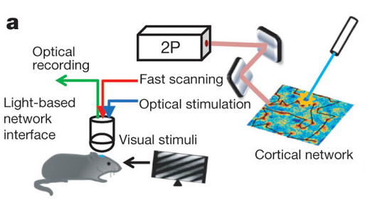
We published another double header yesterday, this time on the role of particular cell types in visual responses. Both studies describe the effect of optogenetically manipulating various interneuron classes in mouse visual cortex. The papers are Lee et al. from Yang Dan‘s lab and Wilson et al. from Mriganka Sur‘s labs. And in fact, both were preceded by Atallah et al. from Massimo Scanziani’s lab, which appeared in Neuron earlier this year. Which means a bonanza of data on the effects of activating parvalbumin-expressing interneurons, and also a bonanza of different conclusions about their exact role – everyone comes to slightly different conclusions.
It’s Friday night now over here, so I’ll leave it at that. But to give you an idea of how much (or little) people publish with us more than once a year, here are wordles showing authors of our neuroscience papers in recent years. As it turns out, an imperfect way of displaying the data (it clearly highlights the general need for a means of disambiguating names in science!) but enjoy anyway (click on images to enlarge).


From Figure 1, Li et al.
I wanted the title of this post to be “A tale of two one two three papers” but I couldn’t figure out how to get strikethroughs in the title field. And I thought “A tale of two, make that one, no make that two again, oops now three” might be a bit cumbersome. As promised, here’s another installment of the discussion of what happens when we receive conceptually related/overlapping papers. It starts with a paper that appeared just yesterday in Neuron by Kenichi Ohki and colleagues describing how mouse visual cortex neurons that developed from the same neural progenitor cell tend to be more similar functionally than those that did not.
Why is this significant? First a little background. Cells in visual cortex are tuned to different aspects of visual stimuli, such as orientation or direction, and anatomically are organized quite specifically. Cells with similar preferences tend to cluster together and to be selectively connected with each other (though to differing degrees in different species), and this specificity may underlie some of the many computations required to turn photons of light hitting our eyes into comprehensible percepts. It’s been proposed that this clustering could start in early development; neurons born from the same neural progenitor migrate vertically to form columns of sibling neurons, and could be the basis for clusters of adult cells with similar properties. That link hasn’t been demonstrated experimentally until now, and Ohtsuki et al. provides some evidence in support of it.
Now, visual cortex aficionados among you may think this sounds a bit similar to Li et al., a paper by Yang Dan and colleagues that appeared a few months ago, and indeed it is. And you may also recall that THAT paper appeared alongside Yu et al. from Songhai Shi’s lab about the development of synapses between sibling neurons.
So here’s the story from the beginning (or rather, the beginning of our involvement with the manuscripts).
We first received Yu et al., which concerns how sibling neurons in cortex preferentially form electrical synapses (gap junctions) early in development and the consequences this early gap junction signaling has for the formation of specific chemical synapses later. “Interesting but thin,” we thought when making our initial assessment to review, “wouldn’t it be nice if the authors could demonstrate some functional consequences?” We then received Li et al. showing just that; they found that sibling neurons are more similar functionally, and if you block gap junctions early in development, that increased similarity goes away. “Interesting” we thought (again) “but a bit thin.” Since one of the authors was on both papers, our next thought was, “Aha! together these would make one amazing paper.” So we approached the reviewers and the authors about the idea of combining the two into a single paper. And we were most politely but firmly dissuaded from the notion. What persuaded us was not just the conceptual advance offered by each individual paper, but also the human factor. These were independent projects done in different labs, with different first authors who deserved credit for their work. We switched our focus to what the reviewers felt they needed to see in order to be persuaded of the robustness of the individual conclusions, and were happy to accept both for publication after some revisions.
This is the point in the story where the Neuron paper enters, the one with highly overlapping conclusions to Li et al. The authors of the Ohtsuki et al. have graciously granted us permission to divulge that their paper was first submitted to us and that we had to decline it (we wouldn’t reveal this information otherwise). Our editorial assessment was that (obviously) the topic of the paper was interesting, and that the work appeared high quality, but that novelty was compromised. Even so, we did consider giving it an expedited review given how close in time it followed, and because its conclusions did differ somewhat. But, Li et al. contained additional parts to the story that Ohtsuki et al did not have, including manipulations to disrupt the sibling cell similarity (ie blocking gap junctions early in development) and additional characterization of multiple visual properties. In the course of our deliberations on the two Nature papers, we had determined that publication of Li et al. was conditional upon the authors strengthening those additional pieces of the story. So we felt it would not be consistent or fair to consider a paper that did not contain them, nor did we think it would be productive to ask the authors of Ohtsuki et al. for months’ more worth of work in this situation. We felt the best we could offer (in our pages) was a Brief Communications Arising once Li et al. had been published. Happily, the authors found another journal able to publish their paper on a relatively fast time frame and I’m glad to be able to present the whole package here today.

From Mrsic-Flogel & Bonhoeffer
If you want to read more about the science, I direct you to not one, but two, excellent commentaries: one in our pages by Tom Mrsic-Flogel and Tobias Bonhoeffer about the two Nature papers and one in Neuron by Gordon Smith and David Fitzpatrick. The Neuron commentary provides a great explanation of the similarities and differences between the studies (and suggests a couple of R01’s worth of future experiments). I highly recommend you check them out.
12/11/12: edited to fix typos – thanks to Steve Esterly for flagging
]]>
From Won et al.
Again, we’re behind on blogging – you guys are keeping us busy with great neuroscience – but here is the story of a pair of papers that appeared back to back in last week’s issue and a continuation of the discussion started here by Noah about the process of joint publication. The two papers by Tobias Boeckers and colleagues and by Eunjoon Kim and colleagues were independently submitted and both describe autism-like phenotypes of mice with mutations in the gene Shank2. In human studies, SHANK2 has been associated with rare cases of autism and these two mice add to the ever-growing list of rodents (according to SFARI.org, 17 rodent models debuted in 2011 alone) that are being created to investigate the functional consequences of genetic mutations linked to autism, in the hopes of understanding mechanisms underlying core symptoms. Shank2 is a scaffolding protein that regulates excitatory synapse function by holding together various molecules such as neurotransmitter receptors and signaling proteins. Mutations in another member of the same gene family, SHANK3, are also associated with human autism, and mutant mice display behaviors reminiscent of ASD symptoms, such as social deficits and obsessive behavior. So this protein family, and more generally, glutamatergic transmission, is potentially one promising line of investigation.
Each of today’s papers describe mice with striking behavior (hyperactivity and social deficits) and alterations in excitatory neuronal activity. And that’s where things start to get (even more) interesting. The mice carry very similar genetic mutations, and have similar behavior, yet are opposite in some aspects of electrophysiology. Boeckers and colleagues report enhanced NMDA receptor-mediated excitatory currents, Kim and colleagues a reduction. Kim also found that restoring NMDAR function pharmacologically improves social deficits, but not other behaviors.
Video: Repetitively jumping Shank2 mouse from Won et al.
A lot of science we could discuss here… but on to editorial issues. Both papers had what one of my former advisors used to call “a difficult birthing process”. Both were rejected at some point, and successfully appealed after inclusion of additional data to strengthen existing conclusions and to extend the studies conceptually. There was discussion of whether the individual components (from molecular to electrophysiology to behavior) of the studies told sufficiently consistent a story. There was discussion of whether the requirement for conceptual advance (i.e. mechanism) needed to raised in light of the existing work on Shank3. And much much more… but obviously, in the end, we agreed that both papers deserved to be published, even though the inconsistent electrophysiology left a question mark. As the papers were not co-submitted, we did not ask that the authors attempt to reconcile the differences. All we could and did do was to seek overlapping reviewers to ascertain, to the best they could tell, that each study was technically sound. Once that had been determined, we did our best to coordinate their appearance by publishing back to back in print (one paper was accepted and published online earlier). In cases where papers ARE formally co-submitted, and therefore we assume all authors are aware of each others’ results, we do sometimes ask that the authors attempt to resolve discrepancies between studies or perform direct comparisons in the interests of advancing the overall scientific picture. One recent example is a trio of exome sequencing papers (coincidentally, also about autism) we published a few months ago. In that case, one of the reviewers requested that the authors compare and cross-reference their results, which they graciously did.
In the case of the Shank2 papers, the papers were highly conceptually related and accepted within weeks of each other, so the case for joint publication was obvious. The situation gets more complicated when papers are at very different stages of the process but that will have to be the topic for another day. Watch this space…
]]>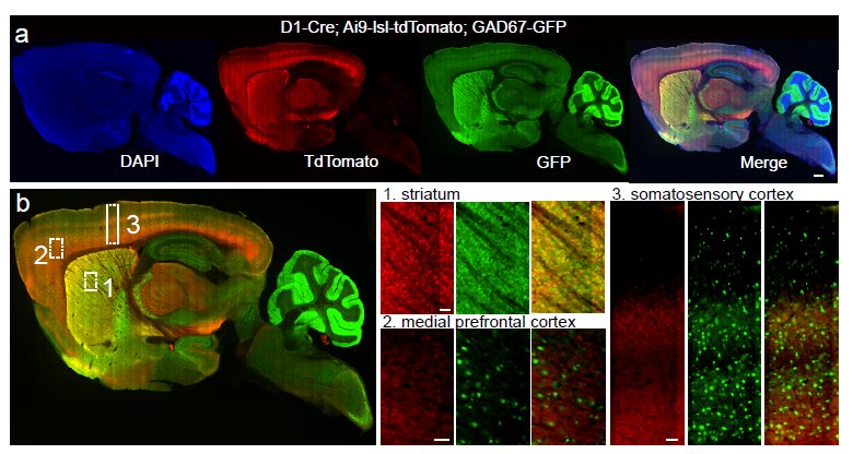
Whole-brain anatomical mapping of D1-Cre expression in inhibitory neurons (from Supp Fig.2)
It really is an embarrassment of riches here at Nature these days, what with so many excellent neuroscience-related studies emerging. Just in the last couple of weeks, we’ve had the following studies:
- Covert learning by a basal ganglia circuit, despite no participating in the behavioral practice.
- Reach and grasp by people with tetraplegia using a neurally-controlled robotic arm.
- A non-transcriptional circadian cycle conserved across all domains of life.
- Identification of mechanisms linking cerebro-vascular integrity to neurodegeneration.
- Metabolic coupling of glia with axons in the long-term maintenance of axonal integrity.
So really, a lot to write about from a science perspective. However, this blog is dedicated to bringing you the editorial back-story, so I wanted to touch on yet another interesting study, published in print today. This new paper offers an opportunity to discuss an important editorial issue: the manuscript appeal process. For more details, you can always read the appropriate section in our guide to authors. But it’s often helpful to follow a particular [successful] example in order to illustrate the process.
Initially, the lab of Bernardo Sabatini submitted the manuscript “Recurrent network activity drives striatal synaptogenesis,” which we found to be interesting since it explored activity-dependent circuit maturation in a brain area other than sensory cortex. They looked at the basal ganglia, which lack the lamination and structure of cortical tissue as well as sensory input. With these nuclei important for not only complex motor behavior, but also reward-based learning, understanding the development of this network is important. Here’s the requisite background:
Excitatory input from the cortex and thalamus enters the basal ganglia through the striatum, where it is locally processed and transformed into two inhibitory, GABAergic outputs called the direct and indirect pathways7. Each pathway arises from a distinct class of spatially intermixed medium spiny neurons (MSNs) with differing projections and molecular characteristics8. These projection patterns suggest opponent effects on basal ganglia output: direct pathway MSNs form synapses in substantia nigra reticulata (SNr), the basal ganglia output nucleus, whereas indirect pathway MSNs form synapses in globus pallidus, which in turn inhibits the SNr9. Because the SNr provides GABAergic inhibition to the thalamus, which subsequently activates cortex through glutamatergic synapses, the interactions of basal ganglia, thalamus and cortex can be simplified as a closed loop, differentially controlled by the direct and indirect pathways (Supplementary Fig. 1). Anatomical evidence supports this model10, 11 and the opponent roles of the two pathways on motor behaviour have been recently demonstrated in adult rodents12, 13. In addition, in vivo recordings and circuit tracing indicate that different corticostriatal inputs are processed through segregated, parallel networks6, 10, 11, 14. Given this organization, establishing correct wiring of the cortex-basal ganglia–thalamus circuitry poses a significant developmental challenge, requiring that functional interactions be maintained over polysynaptic networks comprised of mixed inhibitory and excitatory projections.
So how to probe the maturation of these looped pathways? To make the initial observations, Sabatini and colleagues made transgenic mice incapable of releasing GABA from either the direct or indirect pathway in the basal ganglia during early postnatal development. A technical tour-de-force followed to produce the basic result: silencing of the direct pathway led to hypo-innervation of the stratum by excitatory inputs, while silencing of the indirect pathway resulted in the opposite, excitatory hyper-innervation. Therefore, the balance of activity between the direct and indirect pathways dictated the extent of postnatal excitatory innervation in the striatum. Or, to put it more simply, these data demonstrated how this circuit essentially wired itself, with the output controlling the development of the input.
Initially, the referees were encouraging, but found significant technical deficiencies involving the specificity of the genetic methods, amongst other concerns. All the refs had suggestions for important controls and without them. had dampened enthusiasm for publication. Addressing these concerns would be a significant amount of work, but not beyond the scope of an ambitious researcher or beyond the scope of a paper attempting to make new claims about striatal circuit development. Alone, such concerns would have easily persuaded the editors to invite back a revision. However there were other lingering concerns that together with the technical shortcomings soured the mood on this study. The reviewers also raised general novelty issues, since it is well-known from many brain areas that any manipulation of circuits on a gross level can lead to innervation changes. A somewhat broad damnation, but worth considering nonetheless. This criticism also related to the next, namely that to really make a valuable contribution to the competitive field of circuit development, the authors would need to expand this study further, supplying additional data allowing a better understanding of other components within the circuit. For example, exploring how exactly the corticostriatal inputs influence basal ganglia synaptogenesis. It begged the question: do the authors understand the physiology and timing well enough to predict how their manipulations would affect the striatum, not by disrupting the striatum itself, but through control of the descending cortical circuits? Such an extension would indeed be a strong component to the story, but would also represent a commitment to future experimentation. On balance, we rejected the paper, believing that such an extension would be important for the story to remain editorially viable. Simply fixing the technical concerns would not be enough.
The authors, holding a comprehensive “road map” to publication in hand (the reviews) and knowing that they had the capacity to address all concerns, decided to improve the story based on the referees’ recommendations and contacted me months later to gauge interest in a revised manuscript. They had completed an extensive re-analysis of the genetic specificity of their animals, documented the extent of their experimental system’s “leakiness,” added additional transgenic lines to corroborate their previous conclusions and completed the desired extension. The authors manipulated corticostriatal inputs independently of basal ganglia output and observed the same synaptogenic phenotypes across different developmental time windows. With these new data, the authors “closed the loop” of their circuit, demonstrating the validity of the proposed synaptogenic model. With regards to the novelty criticisms, the authors argued that their story went well beyond “gross manipulations” and that this represented the first study exploring synapse development in a region containing multiple nuclei and intermingled circuits. In addition, unlike most circuit activity manipulation studies in sensory cortex, which often involve large-scale disruptions like eye suture or whisker plucking, this study utilized specific genetic targeting to single-out a neuronal subtype for manipulation. We were (editorially) cool with this updated novelty assessment and argument.
Of course, any consideration of a manuscript without an invitation to resubmit would be considered as an appeal of our original decision. Thus, the authors uploaded a new version of the manuscript, along with a rebuttal to the reviewers and an appeal letter to the editors, explaining why further consideration of the study was merited. In this particular case, the rejection had been (bear with me) a “soft hard-rejection.” Many consider a “soft rejection” one in which the journal invites back a revision. A hard rejection is one without review or one after review in which the editor sees no path to publication, even if technical concerns were satisfied (in other words, the editor screwed up on gauging editorial interest and novelty within that community.) A “soft hard-rejection” would fall in between, when an editor could see a place for the conceptual story within the journal, but feels the journey to that point would be too great or beyond the scope of a single manuscript. In my notes on the first version of this paper, I explicitly conveyed to my colleagues that if an extended story ever did return, we would be wise to re-consider.
So if we liked the story, even just a little, why not simply invite back a revision in the first place? Well science is not done in a vacuum and all too often, the complexities of “real-life” interfere with scientific progress. The lead author has found another job and wants to leave the lab ASAP. The mouse colony was infected with a virus and now all of those precious transgenic animals are in quarantine for three months. Maybe it’s time to just get over that hurdle of publication, dedicating the mental energy and experimental effort to the next project. These aren’t editorial choices for those who work for a journal. These are choices to be made by the lab and authors. In cases where there is an extensive amount of work required, more than perhaps we feel is reasonable (meaning the manuscript is only a more modest advance,) we feel it is important to not instill false hope through an invitation of a revision when there is a strong chance that even the revision will fall short of reviewer demands. This may allow the authors an opportunity to ponder the future of the study, free of a mind clouded by the non-guaranteed prospects of publication that an invited revision might offer. After this sober assessment has been made, if the extra 2-6 months of work (or more??) are embraced, upon completion, the appeal process allows the authors back in the door for reconsideration.
Sabatini’s appeal was granted by the editors and the revision was well-received by the reviewers. The end result is essentially what you see now, in press.
]]>
From Ramsden et al. addendum
The (highly abbreviated) life story of a paper appearing in Nature often goes something like this: ideas are birthed and experiments envisioned. Pilot experiments are run, yielding beautiful preliminary data. Replication and controls are then gathered over the course of months, if not years of hard labor. The paper is written, submitted, and reviewed. A few (two is typical) rounds of review and revision later, it is published (with highly variable degrees of reviewer and editorial unanimity). But this is by no means the end, rather, just a milestone in the evaluation process by the community. In journals, post-publication evaluation has traditionally occurred in the form of peer-reviewed follow-up papers or formal commentary. This may change someday as alternative forms of scientific publishing are explored, but for today we’ll talk about a formal addendum we’re publishing on a 2011 paper by Cathy Price and colleagues and invite you to add to the discussion.
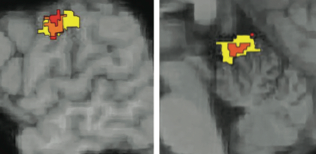
A few months ago, we published the original paper describing structural and functional changes in the teenage brain that accompany changes in IQ over time. The authors measured verbal and nonverbal IQ twice, once in early and once in late adolescence, and also scanned the subjects at both time points. They reported that changes in gray matter density in left motor cortex and anterior cerebellum were correlated with verbal and nonverbal intelligence, respectively. Although structural correlations between brain areas and IQ have been reported before, this paper was unusual in that the authors were able to track the same subjects over time. From the outset, the reviewers were positive about the potential contribution of this study as it not only demonstrated considerable plasticity in IQ during adolescence, but provided evidence of distinct neural substrates for different forms of IQ. Through the course of several rounds of review, various technical issues about statistical analysis, interpretation, and relationship to previous literature were discussed. After publication, we received a formal submission to our Brief Communications Arising section (an online-only section for formal exchanges) raising a concern about statistical inference that was not brought up in the review process. The concern related to the authors’ estimates of how much of the IQ change could be attributed to gray matter changes. The commentary asked whether the reported effect sizes could be potentially distorted by how the analyzed brain areas were selected. Several published commentaries and perspectives have pointed out that using the same dataset to pick out which data to analyze and then analyzing only that dataset can lead to inflated effects. This form of sampling bias, termed a variety of things (including circularity and double-dipping), is common in, but certainly not exclusive to, systems and cognitive neuroscience studies. The concern with this particular paper was that if the relationship between gray matter and IQ was assessed in the MRI voxels selected on the basis of their showing a change with IQ, those effects could be inflated.
We were aware that this was a controversial issue (some of the commentaries created quite a stir when they were published a few years ago). We had heard from many in the community that the practice could potentially invalidate results, but delving more deeply into the issue we realized that there were also other views both on the potential severity of the problem and validity of the practice.
It seemed to us that criticisms should be applied and taken in context and we felt that for this paper, the magnitude of the effects were a key part of the conclusions of the paper – in the field of biological correlates of IQ, the amount of variance accounted for by various factors has been controversial. After consulting with an independent referee (not one of the original reviewers of the paper) we asked the authors to provide a reanalysis that would address the criticism by recalculating their effect sizes using independent data. After several rounds of back and forth between the authors, the reviewer, and ourselves (during the course of which we asked the authors for clarifications of their methods, alternative analyses than the ones they initially provided, and also for an additional analysis to address a different technical concern not raised in the commentary, but flagged by the reviewer) we eventually agreed upon a response that we felt appropriately addressed the concerns. At this point, the authors of the Brief Communications Arising decided to withdraw their commentary.
Nonetheless, we felt that the commentary had raised concerns likely shared by other members of the community (and indeed we had heard informally from others) and that the reanalysis provided clarification of how much the conclusions of the paper were affected by those concerns. We therefore decided to publish the response with a few tweaks to the language as a stand-alone addendum, which you see here today.
A common sentiment we hear regarding the formal corrections/commentary process is that it is frustratingly lengthy and time consuming, which seems anachronistic given the speed and ease with which comments can be communicated on the internet. Indeed, it did take several months to complete this set of communications, due to the many rounds of correspondence and requests for clarification. When the issues are technical and potentially call for revision of published conclusions, it takes time for everyone to present their side of the story and for everything to be evaluated. But to address the need for open communication on a faster time scale, and to host exchanges that we do not feel rise to the level of formal correction, we have an online comments section attached to every paper. At Nature, as with many other journals, these sections are sparsely populated, but they do exist and we want to hear from you!
]]>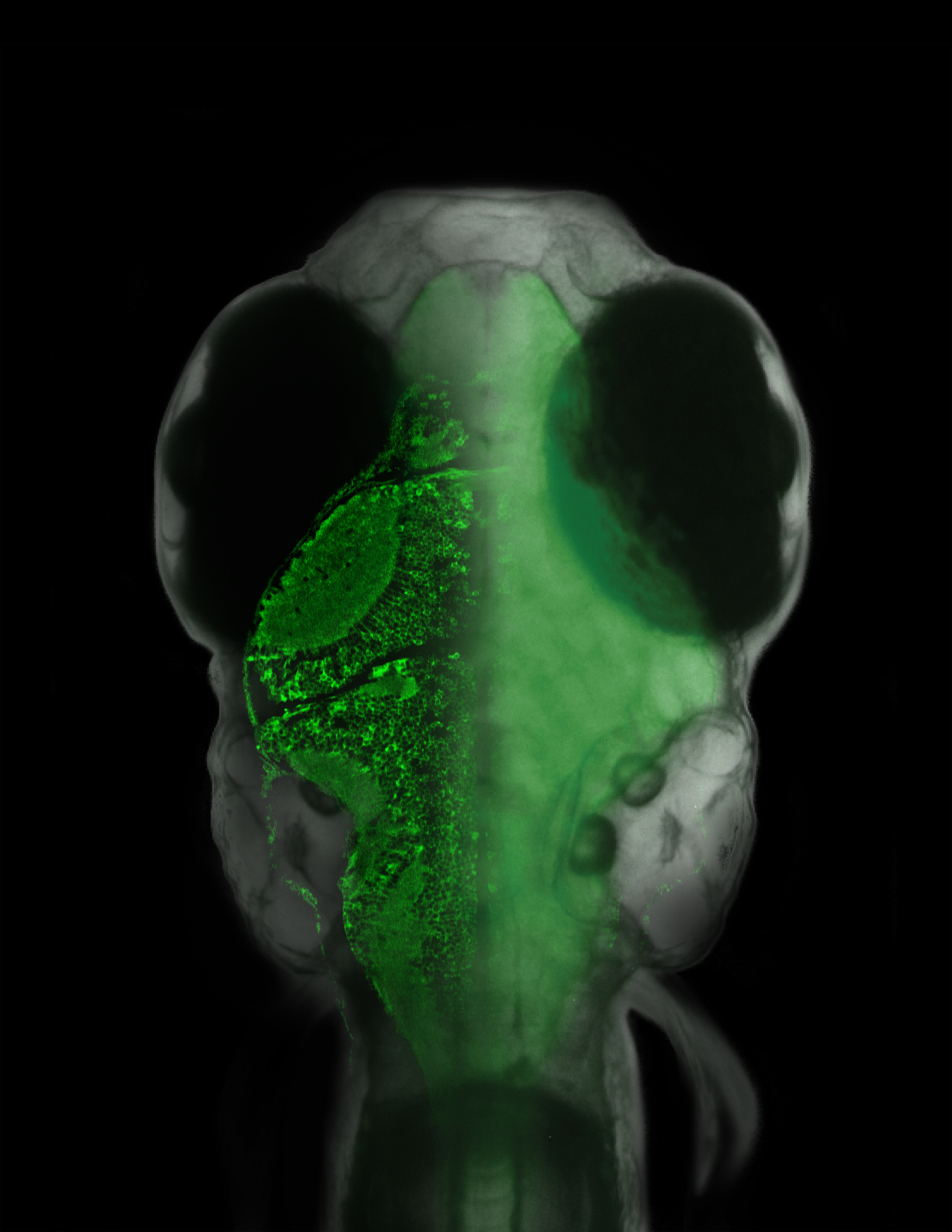
Overlaid on the micrograph of the fish is a slice of its brain measured with a laser scanning microscope, in which single neurons are visible.{credit}(courtesy of Ahrens et al.){/credit}
Sometimes an experiment will just reach off the page and slap you in the face, demanding attention. This happens to me every so often and I must admit, our latest paper from the lab of Florien Engert induced such an experience. There have been several cool, technical tours-de-force (is that proper grammar??) over the last few years involving different creatures navigating in a virtual environment while neuronal activity was monitored. These include a mouse running on a spherical treadmill, as well as a fly marching along a similar treadmill-style ball. But in these examples, having the subject head-fixed (for the stability of recordings in the brain, either with electrodes or through imaging) was moderately non-intrusive since walking motions were independent of the head. The same can’t be said for the subject in this latest example of a virtual reality navigator: a wriggling, swimming fish. Therefore, a more creative solution had to be sought and in a paper published online yesterday, Ahrens, Engert and colleagues decided that paralysis was the way to go in order to follow the neural activity of this navigating fish.
Zebrafish have long been used as a model system for a variety of experiments, but motor control long been at the forefront of topics. They offer many distinct advantages over mammalian-based systems, including the genetic tractability of Drosophila and the ease with which microscopy methods can be utilized; larval zebrafish are transparent, so no surgery is required in order to open up a “window” for fluorescence imaging. And more recently, several labs have begun to develop rich behavioral methods to further explore the limits of probing functional circuitry in these beasts. So what was interesting and cool about this latest fish-based technology? From the authors, here is the system set-up:
To examine neural dynamics across brain areas that drive sensorimotor recalibration, we developed a system to study neural activity at cellular resolution17, 18, using two-photon microscopy19, anywhere in the brain20 during closed-loop optomotor behaviour in larval zebrafish. These animals have a small and transparent brain that is readily accessible for optogenetic recording and stimulation21, 22, electrophysiology23 and single-cell ablation24. To remove motion artefacts25, 26, we developed a swim simulator for completely paralysed larvae (Fig. 1a). Motor commands, or ‘fictive swims’, are recorded at the motor neuron level8, 27, 28 (Fig. 1c, d) and translated, in real time, into visual feedback that resembles the optic flow of freely swimming fish (Methods). This constitutes a fictively driven virtual-reality setup. Simultaneously, a two-photon microscope scanning over a transgenic fish expressing GCaMP2 (ref. 29) in almost all neurons20, 30 allows activity to be monitored throughout the brain at single-neuron resolution. As the experimenter is in complete control of the visual feedback, this allowed us to study neural dynamics during visually guided motor adaptation throughout the brain at the cellular level.
Allow me to translate: “WE CREATED “THE MATRIX” FOR FISH.
Using this system, the authors were able to break down neural response properties during behavioral adaptation to perturbations in the virtual environment into four different categories, and the excellent spatial resolution of this technique allowed them to accurately map the anatomical substrate for each response. Some of these responses were observed while the fish was progressing through behavioral adjustments to the stimulus provided in the virtual world, strongly suggesting that focus on these elements could potentially yield functional insights into adaptive motor learning. Indeed, laser-based lesioning of one identified site, the inferior olive, did negatively affect visually-induced motor adaptation learning. Therefore, the authors had identified a powerful model for the study of brain-wide activity during adaptive locomotion.
The reviews for this paper were quite positive with regards to the innovative technology and each easily recognized the potential for this system, but the original version of the manuscript failed to provide a “proof-of-principle” for its utility; i.e., demonstrating novel insights into motor adaptation programs. Two referees even had concerns that there was a novelty issue when comparing this system to other virtual reality set-ups, including those mentioned earlier. At Nature, we aren’t shy about publishing manuscripts that are wholly-focused on technology and innovation, but in this particular case, although I was dazzled by the technique, we needed to go a little deeper into the biology in order to differentiate this system from the others and demonstrate its power. One could potentially make the argument that working in zebrafish is not as common as working in mammals or even insects, so if this paper was to be significant for a very broad audience, we had to throw some biology at the reader, in addition to the cool kit.
The authors graciously agreed to explore the circuitry further, adding new experiments to test the role and functionality of some of the neural response properties they observed. It took some time to get everything right, but in the end, both editors and (most) reviewers were satisfied that this paper had reached a point where it was not only showcasing the methodology, but also providing some novel biological insights. I say “most” because as is the case often times, not all reviewers were on board with the changes. In a rare occurrence, one reviewer was strongly supportive of publication in the initial round, with this existing more as a methods paper, but soured as the paper became more complex. The “biological insight” observations and extra modeling were not as graciously received, with this reviewer feeling these parts detracted from the rest of the paper. It’s not that this referee undervalues biological insight, rather the main concern was that the new insights require greater development and more sophisticated manipulation beyond lesion-based experiments to provide real value (one can imagine the power of working optogenetics into this set-up.) Fair enough, but in the end we felt the manuscript benefited from this brief, natural extension, to best-demonstrate the power of an exciting new system. Feel free to disagree with this editorial decision in the comments.
]]>
I’m on the road (attending a symposium at MIT: New Insights on Early Life Stress and Mental Health) so this one’s going to be brief. Neural prosthetics are an exciting interface between basic research and technology, an area where the path from fundamental discoveries in the organization and function of the brain to translational advances has been remarkably clear. Cochlear implants have already demonstrated their utility for replacing/enhancing auditory function, and more and more promising advances are coming out all the time in retinal implants. Motor prostheses are another exciting area with the promise to restore motor control to paralyzed individuals and today’s paper by Lee Miller and colleagues represents another step towards a potential prosthetic for spinal injury patients.
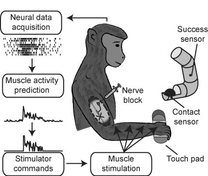
The paper describes an advance in functional electrical stimulation (FES), a procedure by which signals from motor cortex are decoded and used to directly stimulate the arm muscles, bypassing the spinal cord. The subjects in this case are monkeys whose arms are temporarily paralyzed by injecting lidocaine into the peripheral nerves. This work follows on from previous work from both this group and others demonstrating the efficacy of FES for simple movements, and extends it to grasping tasks requiring the coordination of multiple muscles. If a picture says a thousand words, I don’t know how many a movie says, but the video clips associated with the paper explain what the animals could and couldn’t do with the FES better than I ever can:
Examples of Monkey J executing the ball grasp experiment during normal, FES and catch trials.
Monkey T executing the ball grasp experiment during FES and catch trials.
The demonstration is indeed compelling with translational promise, but from an editorial standpoint, these are always interesting papers. As one reviewer once said about neural prosthetic papers (I’m paraphrasing here), you neither have a novel conceptual advance, nor an engineering benchmark to judge them on. Like other translational papers, they fall under a slightly different rulebook, and tend to involve more subjective evaluation than usual. In this case, there was a lot of discussion by the reviewers of how big an achievement the current work represented over the previous FES demonstrations. There was much discussion of whether they had done enough to demonstrate replicability and generality. Bit of trivia: this paper spent the longest time on my desk of any in recent years. The original version was received back in 2009 and was ultimately deemed promising, but too preliminary as the system had only been demonstrated on only one monkey and on one task. We received the next version at the end of last year, and all editors and reviewers were convinced that the extension to two subjects and two tasks made it a technically sound advance. The question of how many subjects we generally require for a study is a discussion for another day (very brief version: we defer to the field for the standards, but in cases where it is appropriate, we consider whether a result merits publication as a proof of principle).
In any case, this study adds to the list of impressive achievements made in recent years in motor prostheses. It’s a prime example of how apparently academic inquiry – basic understanding of the coding and organization of animal motor cortex – is leading to translational advances that are very likely to improve the lives of human patients in the near future. A good example to use when explaining the importance of basic science to the general public.
]]> **PLEASE SEE UPDATES BELOW**
It is commonly believed that distinct mini-networks of neurons, firing together, may be the means by which memories and other conceptual encoding requirements are handled in the brain. However, it is only recently that we have had the tools available to directly test the sufficiency of such a mechanism. Today, a new study in Nature from the lab of Susumu Tonegawa documents the ability to use light as a means to activate distinct subsets of neurons responsible for the encoding of fear memories.
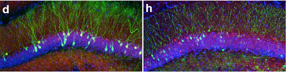
To gain access to cells active during memory encoding, the authors used a clever genetic system in which the promoter of a gene known to be expressed after strong neuronal activation (c-fos) was engineered to drive expression of channelrhodopsin, a light-activated channel able to induce neuronal firing after exposure to blue light. For temporal specificity, the entire system was also under the control of an inducible element, which only allowed the engineered c-fos promoter to operate in the absence of a chemical (doxycycline, or dox,) usually added to the drinking water of the animals. So, for neurons to eventually express channelrhodopsin, they would have to be dox-free and active. There are a variety of controls in the paper to demonstrate the legitimacy of the system and in fact, one example is in the figure above. On the left is the dentate gyrus region of the hippocampus with cells expressing channelrhodopsin tagged with eGFP (green). Upon light exposure, these cells became active and expressed c-fos (immunohistochemical staining, in red.) Although there is additional red that is not overlapping with green signals, almost all green cells contain overlapping red (c-fos) staining. On the right, the dentate gyrus of another animal is shown with cells expressing eGFP only. There are no neurons that should be responding to light and therefore, red c-fos staining simply reflects randomly active neurons. These red cells do not overlap with green cells at any rate greater than chance, demonstrating the strong relationship between neurons expressing channelrhodopsin and c-fos-active neurons (the former should mostly all be a subset of the latter in the presence of light!)
So now the authors had a system in which they could target active neurons and gain access to them again later. In the key experiment, animals were habituated in Context A, with baseline levels of fear quantified by the amount of time the animal spends “freezing.” Freezing is a good measure of fear in rodents because it is an instinctual reaction in the face of vision-driven predators, like birds of prey, looking for movement to cue them on to a potential meal. Of course, animals expressed little freezing behavior in Context A since there was no obvious threat being presented. Animals were then taken off dox and placed in Context B, where they experienced footshocks, presumably triggering the encoding of fear memories related to this negative experience. Although fear memories can be complex, contextual aspects are known to be documented by the hippocampus, one of the areas of the brain encoding information related to location. Thus, in Context B, these hippocampal dentate gyrus cells were becoming strongly activated in an attempt to encode everything about this potentially dangerous context for future reference. And since dox was no longer in the system, these active cells were happily producing channelrhodopsin. After contextual fear training, dox was reintroduced to stop the expression of channelrhodopsin in additional cells unrelated to contextual fear memory simply through random experience and spontaneous activity. Then came the big test. Would the light-driven activation of those cells firing away during the contextual fear encoding be sufficient to induce the fearful behavior of freezing outside the fearful context? Animals were returned to the safe confines of Context A and were allowed to explore in the presence or absence of light activation of channelrhodopsin-expressing cells. Light was introduced via the surgical implantation of a small fiber optic cable directly above the dentate gyrus. Indeed, light-activation of fear-associated, Context B-linked cells induced freezing in neutral Context A, while no freezing was observed in the absence of light exposure. So essentially, the animals were uncontrollably expressing fearful behaviors simply because circuits created to respond to an experienced dangerous context were being artificially activated, bypassing executive control and simply short-circuiting the initiation of freezing behavioral programs. It’s hard not to think that’s pretty cool.
But despite demonstrating the sufficiency of these microcircuits, or “memory engrams,” to invoke fear response out of context, what would be really nice is also demonstrating necessity. In fact, this was raised during the review process. A subset of the reviewers (and me) would have liked to see the authors disrupt this memory engram in the appropriate context, say by expressing a different light-activated channel in the participating neurons to shut the cells off, and document the result. In this scenario, although the animals would usually invoke freezing behaviors naturally in the dangerous Context B, if the same cells “remembering” Context B were inhibited, then the rodent may not express fear. It sounds great in principle, but the authors basically argued that fear memories are much more complicated than that. Removing only one trigger may not be enough, or perhaps the engram could be more complicated than what they could target genetically for inactivation, so a negative result may not be interpretable. After rounds of discussion and additional controls, both the editors and (most of) the reviewers were convinced that pursuing the question of necessity was much more complex, may not lead to a clean resolution and would unduly delay publication of this exciting result that already demonstrated sufficiency of the engram to induce fear.
This was a good thing for the authors for a number of reasons, not least because after we accepted their manuscript, they were made aware of a similar paper close to publication in Science asking similar types of questions, but using distinct methods and strategies. Now usually we are moderately speedy with preparing papers for online publication (perhaps 3-4 weeks,) but the authors were concerned enough about being “scooped” that we decided to “fast-track” the production of the paper in order for it to appear at the same time as the competing work in Science. It took about a week after receiving the final version from the authors for our production team to create the “near-final version” of the manuscript, the one that is currently linked-to above. Once it was clear that we were on track and ready for online publication, the only thing left to do was to coordinate publication with Science. Simple communications between Nature‘s press office and their counter-part at Science confirmed the presence of a similar paper and determined that Science would be releasing their paper today. So we did the same, with the same exact release time. This paper, “Generation of a Synthetic Memory Trace,” from the laboratory of Mark Mayford can be found here. I would’ve written that one up too, but it’s already past 16:00, I’ve got even more awesome papers to read and I need more coffee.
03/23/12 UPDATE: Your prayers have been answered because Jason Snyder of the Functional Neurogenesis blog has written up more on the complementary paper from the Mayford group. Even more coverage from science writer Ed Yong as well as MIT News.
03/23/12 UPDATE #2: As mentioned in the comments below, Hysell Oviedo remembers the original public demonstration of using this methodological strategy to re-activate specific memory traces using optogenetics. This occurred in a presentation at the Society for Neuroscience meeting in the fall of 2009, from the laboratory of Michael Häusser. Dr. Häusser was kind enough to provide us with the original press release from SfN here [PDF]. This is interesting, given the current discussion over the future of science communication and publications. Under a different scientific culture and data presentation system, this original abstract would have been discussed and vetted for years, inspiring the authors to improve the work in a public manner, with no question as to precedence. Under our current system, unless you were at SfN 09 (and even if you were,) it is likely you never saw this work at all. At least until now…
]]>